
ORTHOPEDICS AND TRAUMATOLOGY
Orthopedics studies congenital and acquired diseases of the musculoskeletal system, and traumatology — damage of bones, joints, muscles, ligaments. An orthopedic traumatologist is a specialist who is engaged in the prevention, diagnosis and treatment of various disorders in the musculoskeletal system associated with both diseases and injuries. Modern departments of medical institutions of orthopedics and traumatology combined advanced diagnostic equipment, minimally invasive surgical technologies, comfort of postoperative stay and round-the-clock monitoring of experienced rehabilitologists.

LIST OF DISEASES IN THE FIELD OF ORTHOPEDICS
- diseases of joints of the limbs and spine (arthritis, arthrosis, bursitis, osteochondrosis, intervertebral hernias, scoliosis)
- various deformities of the foot, fingers and toes, limbs in general, defects in the structure of the thorax
- congenital dysplasia, hip dislocation
- bone diseases, inflammation of tendons, ligaments, muscles
- torticollis
- injuries — bruises, dislocations of joints, sprains and ruptures of ligaments, ruptures of muscles and tendons, bone fractures, damage to the spine, head injuries, as well as their complications

LIST OF DISEASES IN THE FIELD OF ORTHOPEDICS
- diseases of joints of the limbs and spine (arthritis, arthrosis, bursitis, osteochondrosis, intervertebral hernias, scoliosis)
- various deformities of the foot, fingers and toes, limbs in general, defects in the structure of the thorax
- congenital dysplasia, hip dislocation
- bone diseases, inflammation of tendons, ligaments, muscles
- torticollis
- injuries — bruises, dislocations of joints, sprains and ruptures of ligaments, ruptures of muscles and tendons, bone fractures, damage to the spine, head injuries, as well as their complications
LIST OF DISEASES IN THE FIELD OF ORTHOPEDICS
- diseases of joints of the limbs and spine (arthritis, arthrosis, bursitis, osteochondrosis, intervertebral hernias, scoliosis)
- various deformities of the foot, fingers and toes, limbs in general, defects in the structure of the thorax
- congenital dysplasia, hip dislocation
- bone diseases, inflammation of tendons, ligaments, muscles
- torticollis
- injuries — bruises, dislocations of joints, sprains and ruptures of ligaments, ruptures of muscles and tendons, bone fractures, damage to the spine, head injuries, as well as their complications
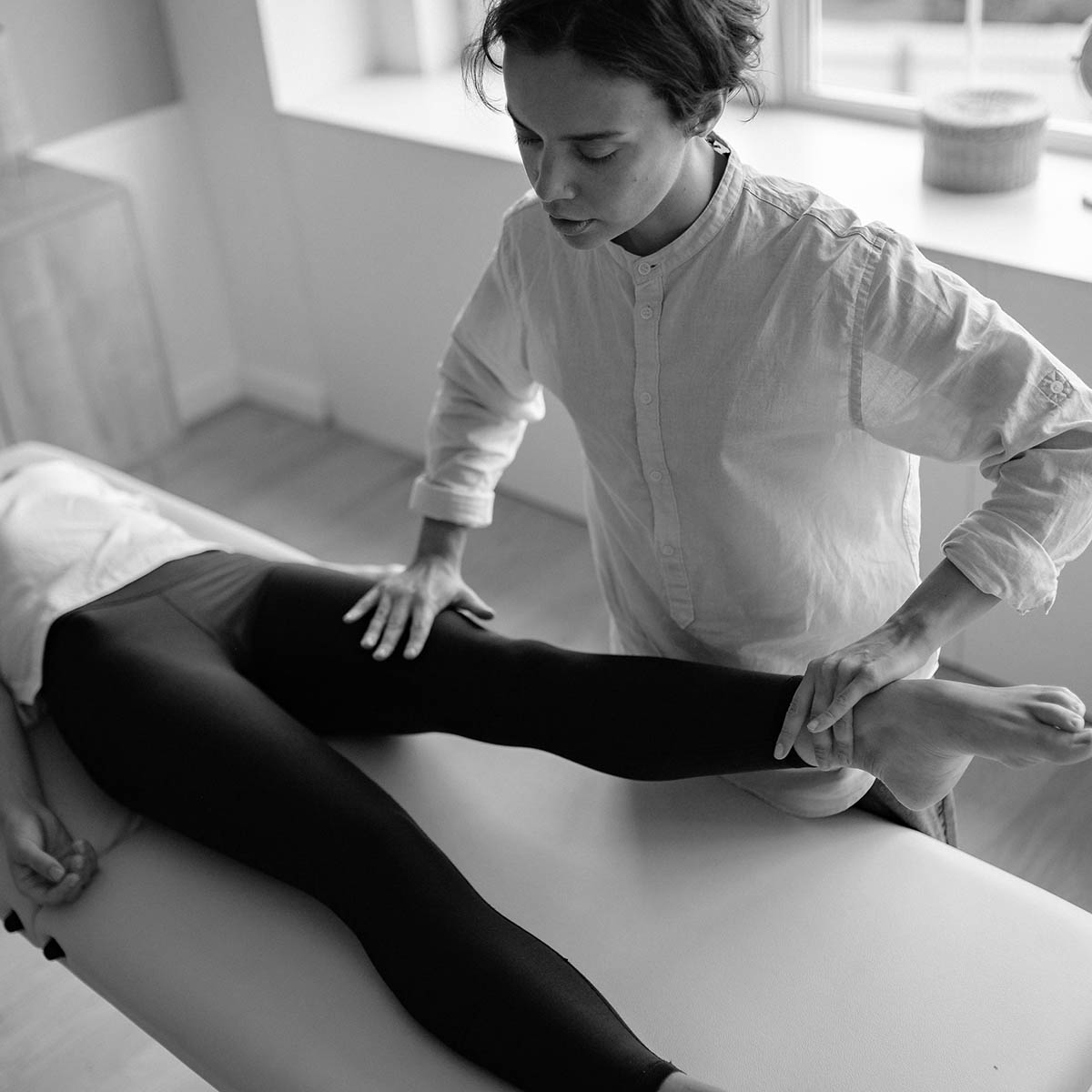
YOU SHOULD APPLY IF YOU HAVE SUCH SYMPTOMS
- even minor physical activities are accompanied by painful sensations in the muscles, quickly leading to fatigue;
- joint mobility is limited, there is a feeling of stiffness;
- tissues around the joints swell, turn red, become hot and painful;
- joints ache at rest or when moving, movements are accompanied by a crunch;
- joint jamming occurs, which is accompanied by sharp pain;
- the joint is deformed, there are differences in the shape of paired, symmetrical joints;
- bones often ache and hurt ;
- you feel numbness of your limbs;
- incorrect posture has been formed or is forming;
- walking is accompanied by limping, strong swinging of the body;
- the length of the left and right arms (legs) varies.
YOU SHOULD APPLY IF YOU HAVE SUCH SYMPTOMS
- even minor physical activities are accompanied by painful sensations in the muscles, quickly leading to fatigue;
- joint mobility is limited, there is a feeling of stiffness;
- tissues around the joints swell, turn red, become hot and painful;
- joints ache at rest or when moving, movements are accompanied by a crunch;
- joint jamming occurs, which is accompanied by sharp pain;
- the joint is deformed, there are differences in the shape of paired, symmetrical joints;
- bones often ache and hurt ;
- you feel numbness of your limbs;
- incorrect posture has been formed or is forming;
- walking is accompanied by limping, strong swinging of the body;
- the length of the left and right arms (legs) varies.
ARTHROSCOPY OF THE SHOULDER JOINT
The shoulder joint is the most mobile in the human body. Its spherical shape allows a wide range of movements of the upper limb. However, this advantage causes its high vulnerability, so the list of possible damage to the shoulder joint is extremely wide.
The limb moves thanks to the joint apparatus. This is a rather complex structure, and damage to any of its components: muscles, ligaments, cartilage or others, causes painful problems and limitations of joint activity.
- Arthroscopy of the shoulder joint is a modern and safe method of surgical intervention. It is used for diagnostic and therapeutic purposes in case of:
- injuries of the rotator (rotator) cuff tendon, articular lip, long bicep head;
- chondromatosis — the presence of free chondromatous bodies inside the joint that occur due to the degeneration of the synovial membrane;
- congenital disorders of the anatomical structure of the shoulder joint.
The result of the intervention is influenced by a number of factors related to the patient’s age, anatomical features of the body, the trauma itself. The competence of an orthopedic traumatologist and conscientious implementation of medical recommendations by a patient also play a significant role. Subject to the rules of the rehabilitation period, motor activity in young people, as a rule, returns in full.
Most of the patients are athletes and young people having traumas. And also elderly people with degenerative changes in the joint. The presence of symptoms does not always mean the necessity of intervention. Some patients should be prescribed conservative therapy.
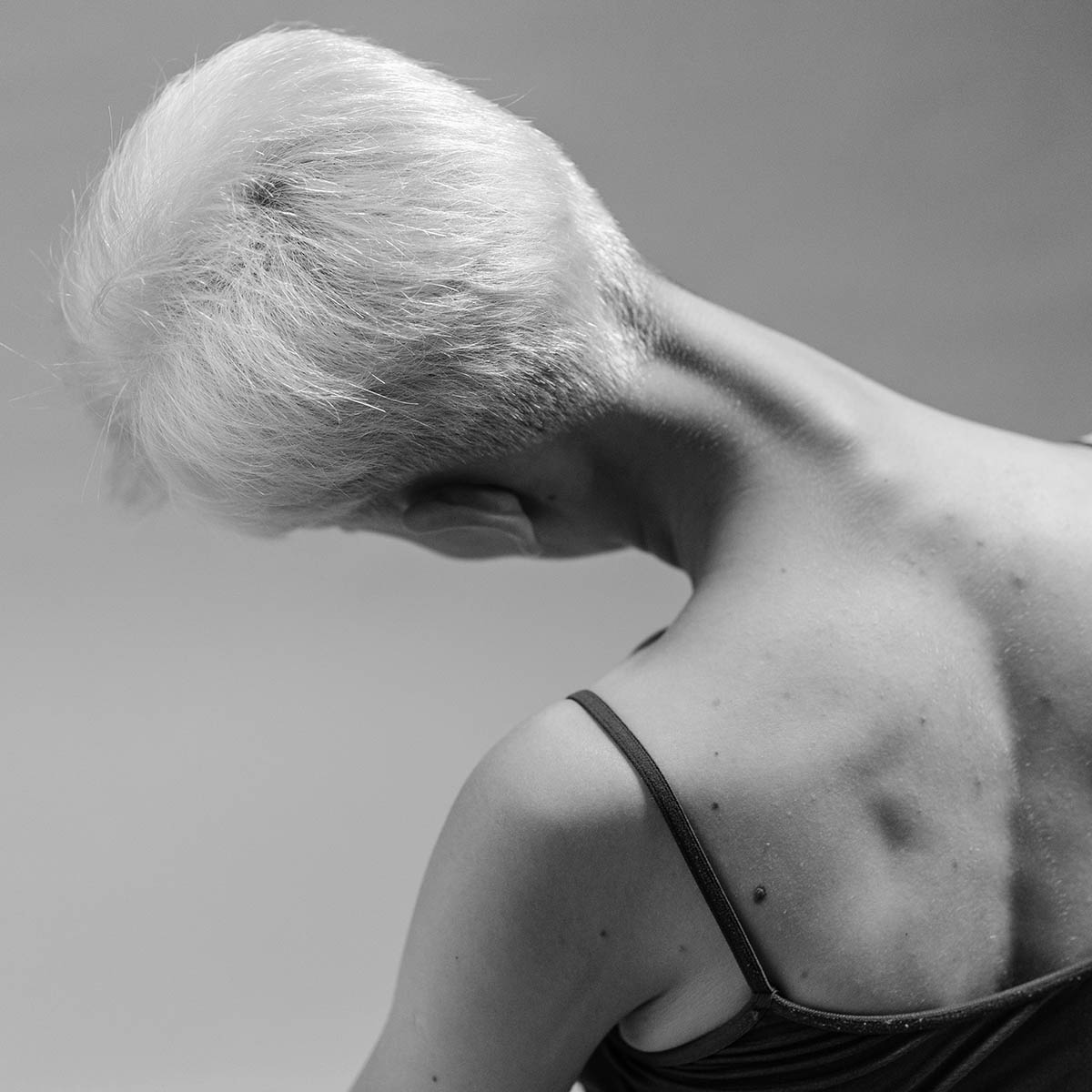
ARTHROSCOPY OF THE SHOULDER JOINT
The shoulder joint is the most mobile in the human body. Its spherical shape allows a wide range of movements of the upper limb. However, this advantage causes its high vulnerability, so the list of possible damage to the shoulder joint is extremely wide.
The limb moves thanks to the joint apparatus. This is a rather complex structure, and damage to any of its components: muscles, ligaments, cartilage or others, causes painful problems and limitations of joint activity.
- Arthroscopy of the shoulder joint is a modern and safe method of surgical intervention. It is used for diagnostic and therapeutic purposes in case of:
- injuries of the rotator (rotator) cuff tendon, articular lip, long bicep head;
- chondromatosis — the presence of free chondromatous bodies inside the joint that occur due to the degeneration of the synovial membrane;
- congenital disorders of the anatomical structure of the shoulder joint.
The result of the intervention is influenced by a number of factors related to the patient’s age, anatomical features of the body, the trauma itself. The competence of an orthopedic traumatologist and conscientious implementation of medical recommendations by a patient also play a significant role. Subject to the rules of the rehabilitation period, motor activity in young people, as a rule, returns in full.
Most of the patients are athletes and young people having traumas. And also elderly people with degenerative changes in the joint. The presence of symptoms does not always mean the necessity of intervention. Some patients should be prescribed conservative therapy.
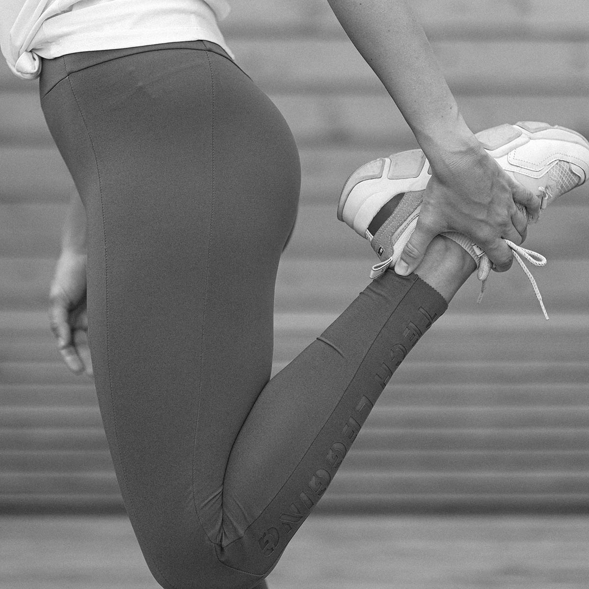
ARTHROSCOPY OF THE KNEE JOINT
Knee arthroscopy is a minimally invasive surgical intervention in the knee joint area for diagnostic or therapeutic purposes. The operation is carried out with minimal tissue trauma and guarantees rapid recovery of joint functions.
Arthroscopy is performed without incisions, but with the help of small punctures. Through punctures, an arthroscope is introduced — a 4-5 mm tube on which a lens system is located. This allows an orthopedic traumatologist to examine the joint with multiple magnification, assess the condition of all its structures and eliminate the damage.
The knee joint is one of the largest in the human body, subject to heavy loads, so it is often injured. The most frequent symptoms for injuries or diseases of the knee: pain, swelling, deformation and inability to move the leg. In this case, do not wait — you need specialist advice.
Knee arthroscopy is performed to diagnose or treat the following knee joint pathologies:
- knee injury,
- arthrosis or arthritis,
- ruptures or damage to the anterior and/or posterior cruciate ligament,
- dislocation or instability of the patella,
- damage to the synovial membrane,
- ageing of cartilage tissue leading to cartilage destruction,
- meniscus cyst,
- chronic inflammation in the joint area,
- the presence of excess fluid, mamelons, bone fragments in the joint.
Before performing arthroscopy of the knee joint, it is necessary to take a number of tests in order to identify concomitant pathologies. Arthroscopy can only be performed by a highly qualified specialist.
ARTHROSCOPY OF THE KNEE JOINT
Knee arthroscopy is a minimally invasive surgical intervention in the knee joint area for diagnostic or therapeutic purposes. The operation is carried out with minimal tissue trauma and guarantees rapid recovery of joint functions.
Arthroscopy is performed without incisions, but with the help of small punctures. Through punctures, an arthroscope is introduced — a 4-5 mm tube on which a lens system is located. This allows an orthopedic traumatologist to examine the joint with multiple magnification, assess the condition of all its structures and eliminate the damage.
The knee joint is one of the largest in the human body, subject to heavy loads, so it is often injured. The most frequent symptoms for injuries or diseases of the knee: pain, swelling, deformation and inability to move the leg. In this case, do not wait — you need specialist advice.
Knee arthroscopy is performed to diagnose or treat the following knee joint pathologies:
- knee injury,
- arthrosis or arthritis,
- ruptures or damage to the anterior and/or posterior cruciate ligament,
- dislocation or instability of the patella,
- damage to the synovial membrane,
- ageing of cartilage tissue leading to cartilage destruction,
- meniscus cyst,
- chronic inflammation in the joint area,
- the presence of excess fluid, mamelons, bone fragments in the joint.
Before performing arthroscopy of the knee joint, it is necessary to take a number of tests in order to identify concomitant pathologies. Arthroscopy can only be performed by a highly qualified specialist.
DEFORMATION OF FEET
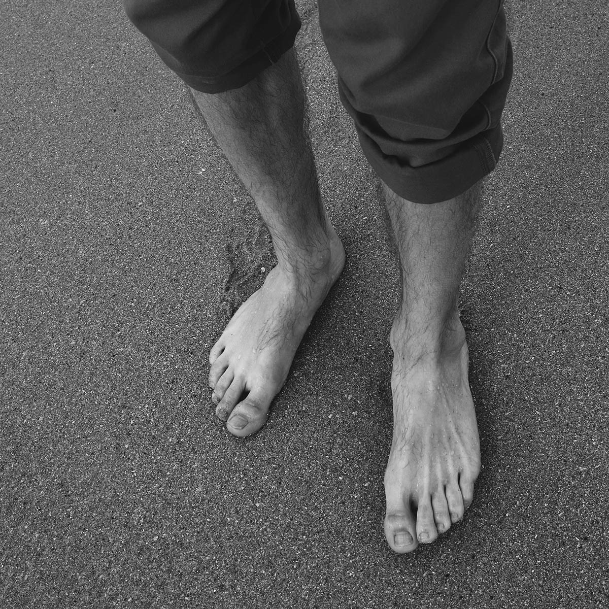
Foot deformity is a change in appearance due to deformation of the bones, and a foot has 26 bones. This becomes noticeable when the length of some bones changes: the bone begins to bulge outward, and the foot itself looks curved. Because of this, discomfort appears and prevents a person from walking and needs treatment.
Foot deformity — a common disease and its causes are diverse:
- Even minor changes in the appearance of the cuts or any external injuries
- paralysis
- uncomfortable shoes or frequent usage of high heels
- being overweight
- lack of vitamins in the body, and especially, poor absorption of calcium
- sedentary lifestyle
- congenital pathology
foot can contribute to the development of other serious diseases or manifest symptoms of diseases, for example, curvature of the spine, various arthrosis and osteochondrosis.
There are 4 stages of deformation of the foot. Treatment is chosen depending on the current stage. The higher it is, the harder the treatment and recovery is for the patient. Often stages are defined in degrees of deviation. Similar to the fight against any disease, it is important to consult a doctor in time to prevent complications.
Treatment of foot deformity is carried out by an orthopedic traumatologist. Each case requires a special approach, especially if we take into account the fact that not only adults, but also children suffer from this. Conservative treatment often only helps to relieve symptoms, but not to remove the real source of the problem. In most cases, surgery is necessary.
The recovery of motor functions and the duration of rehabilitation directly depend on the professionalism of the orthopedist-traumatologist. It is necessary to make the correct diagnosis when a foot is deformed.
DEFORMATION OF FEET
Foot deformity is a change in appearance due to deformation of the bones, and a foot has 26 bones. This becomes noticeable when the length of some bones changes: the bone begins to bulge outward, and the foot itself looks curved. Because of this, discomfort appears and prevents a person from walking and needs treatment.
Foot deformity — a common disease and its causes are diverse:
- Even minor changes in the appearance of the cuts or any external injuries
- paralysis
- uncomfortable shoes or frequent usage of high heels
- being overweight
- lack of vitamins in the body, and especially, poor absorption of calcium
- sedentary lifestyle
- congenital pathology
foot can contribute to the development of other serious diseases or manifest symptoms of diseases, for example, curvature of the spine, various arthrosis and osteochondrosis.
There are 4 stages of deformation of the foot. Treatment is chosen depending on the current stage. The higher it is, the harder the treatment and recovery is for the patient. Often stages are defined in degrees of deviation. Similar to the fight against any disease, it is important to consult a doctor in time to prevent complications.
Treatment of foot deformity is carried out by an orthopedic traumatologist. Each case requires a special approach, especially if we take into account the fact that not only adults, but also children suffer from this. Conservative treatment often only helps to relieve symptoms, but not to remove the real source of the problem. In most cases, surgery is necessary.
The recovery of motor functions and the duration of rehabilitation directly depend on the professionalism of the orthopedist-traumatologist. It is necessary to make the correct diagnosis when a foot is deformed.
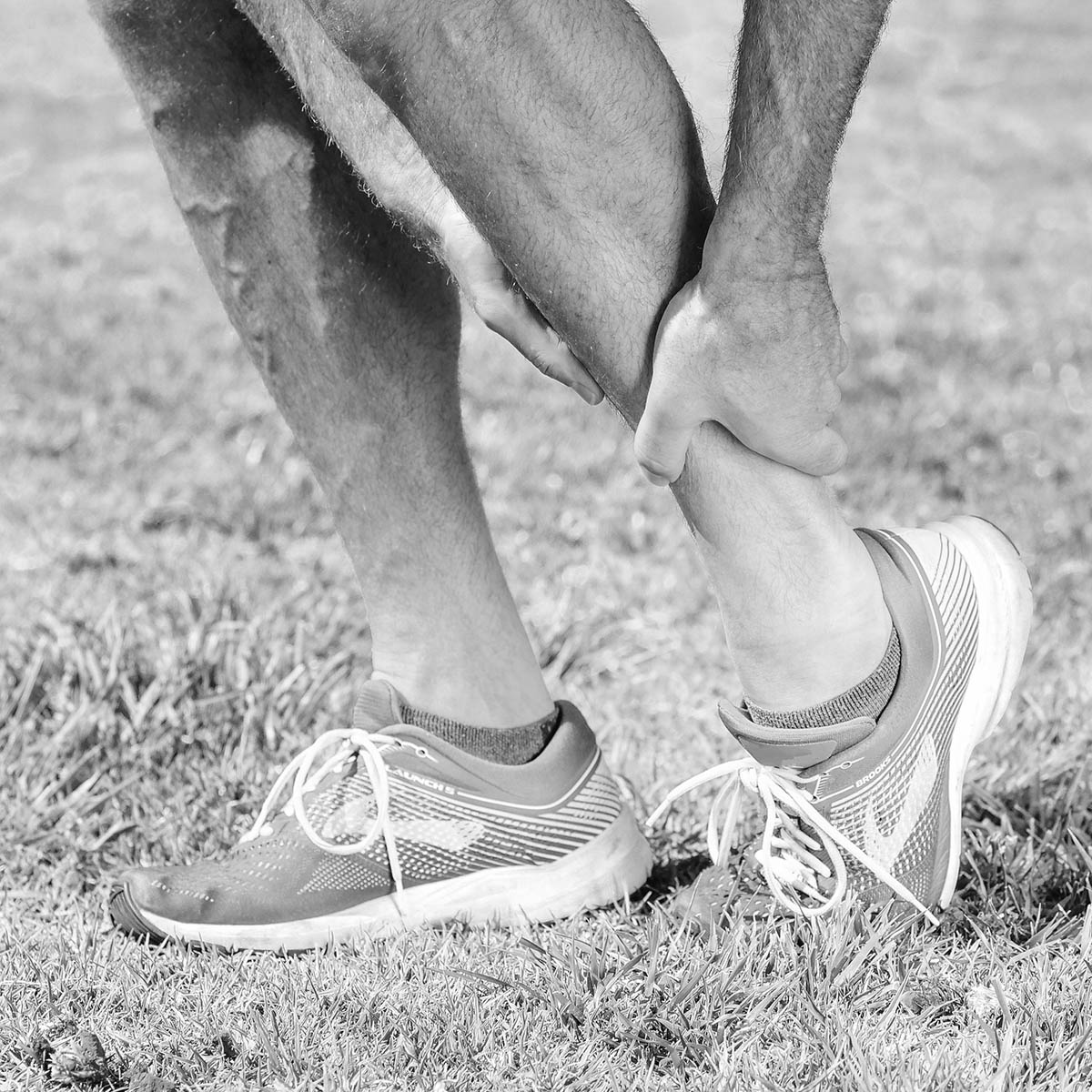
TREATMENT OF FRACTURES
A fracture is a bone injury in which its integrity is violated. This type of injury can occur for various reasons: an accident, during doing sports, strong hits during falls, etc. The timely provision of medical care plays an important role in case of fracture. The main goal in the treatment of fractures of any kind: from finger fractures to fractures of the femoral neck and fractures of the spine is the restoration of the anatomical shape of the bone, the fusion of bone and the restoration of impaired functions.
Depending on the damage, fractures can be classified into:
- closed — the skin is intact
- open — with concomitant wound, rupture of soft tissues and skin
- conditionally open — bone fracture not at the time of injury
Doctors of clinical specialized institutions from Prestige Medical portfolio use the following methods of treatment of bone fractures:
- Conservative method of fixing the damaged bone with a plaster or polymer bandage or fixators. This type of treatment can be used to different types of damage.
- Surgical treatment (metal osteosynthesis or endoprosthetics) — fixing the bone to restore its integrity with the help of implants, pins, plates, screws, anchors.
- External use of metal structures for bone fusion, use devices (spokes, pins, modular, combined).
Traumatological operations, installation of implants and metal structures must be carried out only by experienced specialists, taking into account the individual characteristics of each patient. During the procedure, all parameters of the patient’s vital activity should be monitored, which helps to quickly eliminate complications and respond to unforeseen situations.
TREATMENT OF FRACTURES
A fracture is a bone injury in which its integrity is violated. This type of injury can occur for various reasons: an accident, during doing sports, strong hits during falls, etc. The timely provision of medical care plays an important role in case of fracture. The main goal in the treatment of fractures of any kind: from finger fractures to fractures of the femoral neck and fractures of the spine is the restoration of the anatomical shape of the bone, the fusion of bone and the restoration of impaired functions.
Depending on the damage, fractures can be classified into:
- closed — the skin is intact
- open — with concomitant wound, rupture of soft tissues and skin
- conditionally open — bone fracture not at the time of injury
Doctors of clinical specialized institutions from Prestige Medical portfolio use the following methods of treatment of bone fractures:
- Conservative method of fixing the damaged bone with a plaster or polymer bandage or fixators. This type of treatment can be used to different types of damage.
- Surgical treatment (metal osteosynthesis or endoprosthetics) — fixing the bone to restore its integrity with the help of implants, pins, plates, screws, anchors.
- External use of metal structures for bone fusion, use devices (spokes, pins, modular, combined).
Traumatological operations, installation of implants and metal structures must be carried out only by experienced specialists, taking into account the individual characteristics of each patient. During the procedure, all parameters of the patient’s vital activity should be monitored, which helps to quickly eliminate complications and respond to unforeseen situations.
ADVANTAGES OF ARTHROSCOPY
- preserving tissue integrity
- nice cosmetic effect (invisible scars)
- short period of hospital stay
- significantly less pain compared to open surgery
- low probability of complications
- quick recovery of limb function and return to the usual lifestyle
The decision on treatment is made with the doctor on the basis of diagnosis, patient’s history and his desires as to the future.
ADVANTAGES OF ARTHROSCOPY
- preserving tissue integrity
- nice cosmetic effect (invisible scars)
- short period of hospital stay
- significantly less pain compared to open surgery
- low probability of complications
- quick recovery of limb function and return to the usual lifestyle
The decision on treatment is made with the doctor on the basis of diagnosis, patient’s history and his desires as to the future.
ADVANTAGES OF ARTHROSCOPY
- preserving tissue integrity
- nice cosmetic effect (invisible scars)
- short period of hospital stay
- significantly less pain compared to open surgery
- low probability of complications
- quick recovery of limb function and return to the usual lifestyle
The decision on treatment is made with the doctor on the basis of diagnosis, patient’s history and his desires as to the future.
IS THERE A POSSIBILITY OF A RELAPSE AFTER SURGERY?
The result of the intervention is influenced by a number of factors related to the patient’s age, anatomical features of the body, the trauma itself. A significant role is also played by the competence of an orthopedic traumatologist and thorough implementation of medical recommendations by the patient. Obeying the rules of the rehabilitation period, motor activity in young people as a rule returns in full. Elderly patients can at least perform basic movements, for example, brush their teeth, comb, eat, visit the restroom, serve themselves and so on.
PORTFOLIO OF DIAGNOSTICS FROM PRESTIGE MEDICAL
- Magnetic resonance imaging is an informative instrumental diagnostic method
- Computed tomography as an indispensable method for accurate diagnosis of joint and bone pathology
- Digital radiography. The main diagnostic method for an orthopedist-traumatologist due to the informative value of injuries, diseases, pathologies of bones and joints.
- Ultrasound helps to assess the morphological picture and functional state of the joints with the highest accuracy.
The efficiency and effectiveness of treatment and the full recovery of your health depends the professionalism of the traumatologist-orthopedist so do not hesitate and contact the consulting service of our company.